|
Real-time Lung Radiotherapy Visualization
In this project, we are developing advanced radiotherapy monitoring methods that incorporate physics-
and physiology-based 3D lung dynamics for predicting the amount of radiation dose delivered in vivo
on a lung tumor and its surrounding tissues.
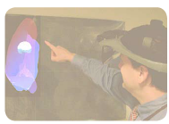
This project focuses on modeling and visualizing the radiotherapy
treatment dose accumulated on a 3D lung tumor, which moves
during breathing. The project aims to address two key issues of
lung radiotherapy, namely (1) to calculate the amount of dose
delivered on a moving lung tumor, and (2) to calculate the
amount of dose delivered on the normal lung tissues that get
exposed to radiation because of the tumor motion. The tumor
motion is coupled with the deformable lung surface model to
simulate the tumor motion for different breathing conditions.
Our main collaborators on this project include METI
Corporation, Columbia University Medical School, John
Hopkins Medical School, The University of Florida,
Washington University Medical School in Saint Louis, The MD
Anderson Cancer Center in Orlando, Ocala Regional Hospital,
The Medical Imaging Group of the Ecole National Superieure
des Telecommunications in Paris.
Additional References
Cali Fidopiastis, User-Centered Virtual Environment Assessment and Design for Cognitive Rehabilitation Applications,
Ph.D. Dissertation, University of Central Florida (2006).
Anand Santhanam, Modeling and Simulation of 3D lung Dynamics,
Ph.D. Dissertation, University of Central Florida
(2006).
Long K. Nguyen, Direct Manipulation of Virtual Objects,
Ph.D. Dissertation, University of Central Florida
(2009).
|